Fluorescence Microscopy
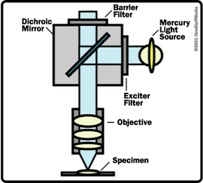
A fluorescence microscope uses a mercury or xenon lamp to produce ultraviolet light. The light comes into the microscope and hits a dichroic mirror -- a mirror that reflects one range of wavelengths and allows another range to pass through. The dichroic mirror reflects the ultraviolet light up to the specimen. The ultraviolet light excites fluorescence within molecules in the specimen. The objective lens collects the fluorescent-wavelength light produced. This fluorescent light passes through the dichroic mirror and a barrier filter (that eliminates wavelengths other than fluorescent), making it to the eyepiece to form the image.
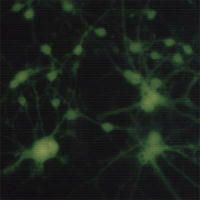
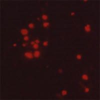
The fluorescent molecules within the specimen can either occur naturally or be introduced. For example, you can stain cells with a dye called calcein/AM. By itself, this dye is not fluorescent. The AM portion of the molecule hides a portion of the calcein molecule that binds calcium, which is fluorescent. When you mix the calcein/AM with the solution bathing the cells, the dye crosses into the cell. Living cells have an enzyme that removes the AM portion, traps the calcein within the cell and allows the calcein to bind calcium so that it fluoresces green under ultraviolet light. Dead cells no longer have this enzyme. Therefore, living cells will fluoresce green, and dead cells will not fluoresce. You can see the dead cells in the same specimen if you mix in another dye called propidium iodide, which only penetrates the dead cells. Propidium iodide binds to DNA in the nucleus and fluoresces red under ultraviolet light. This double-dye technique is used in toxicology studies to determine the percent of a cell population that is killed when treated with some environmental chemical, such as a pesticide.
Advertisement
Fluorescence-microscopy techniques are useful for seeing structures and measuring physiological and biochemical events in living cells. Various fluorescent indicators are available to study many physiologically important chemicals such as DNA, calcium, magnesium, sodium, pH and enzymes. In addition, antibodies that are specific to various biological molecules can be chemically bound to fluorescent molecules and used to stain specific structures within cells. See Molecular Expressions: Fluorescence Microscopy for details and more examples.
In the next section, we'll examine the components of a light microscope and their functions.