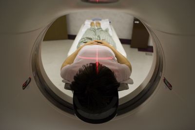
MRI machines use radio waves and magnets to provide an unparalleled view inside the human body. See what it is like to get a head scan on the next page.
Advertisement
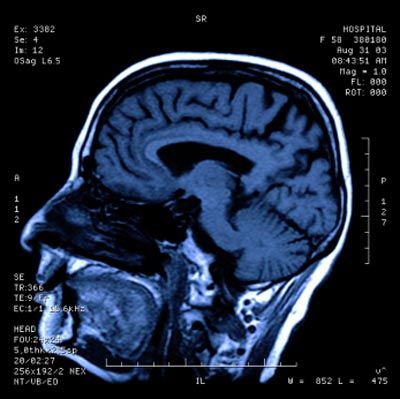
The MRI system goes through the patient's body point by point, building up a 2-D or 3-D map of tissue types to create images. Ever wonder what your brain looks like? See the next picture and find out.
MRI scans can be used to help surgeons accurately locate structures within a patient's brain, in addition to tumors, as seen on the next page.
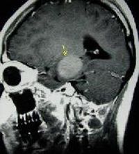
This image shows a tumor growth in a female's brain. MRI systems can distinguish between blood flow and tissue by using a contrast injection. See how injection dyes create an image of the human spine next.
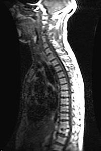
The injectable contrast, or dyes, used with MRI alter the local magnetic field. Next, take a look at internal organs through MRI.
Advertisement
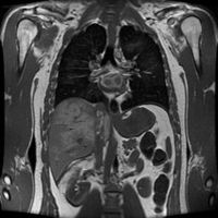
This MRI image shows some of the internal organs in someone's upper torso. An MRI can also be useful in showing broken bones, like on the next page.
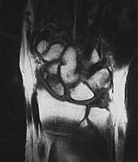
In this MRI scan, you can clearly see the shattered fragments of a human wrist broken from a fall. Next, see what doctors are doing while a scan is taking place.
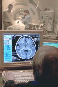
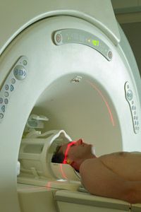
Prior to allowing a patient or support staff member into the scan room, he or she is thoroughly screened for metal objects because of the powerful magnets. See why your doctor might order an MRI scan on the next page.

MRI is ideal for diagnosing multiple sclerosis, tumors, brain infections, torn ligaments, shoulder injuries, bone tumors, cysts or even strokes in their earliest stages. See the future of MRI next.
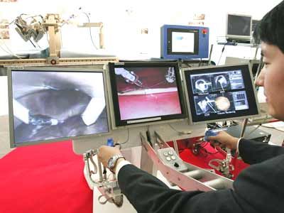
A man demonstrates Hitachi's prototype robot, an MRI image-guided surgical robotic system for caparoscopic surgery. See How MRI Works to learn more.
Advertisement