Image Quality
When you look at a specimen using a microscope, the quality of the image you see is assessed by the following:
- Brightness - How light or dark is the image? Brightness is related to the illumination system and can be changed by changing the voltage to the lamp (rheostat) and adjusting the condenser and diaphragm/pinhole apertures. Brightness is also related to the numerical aperture of the objective lens (the larger the numerical aperture, the brighter the image).
- Focus - Is the image blurry or well-defined? Focus is related to focal length and can be controlled with the focus knobs. The thickness of the cover glass on the specimen slide can also affect your ability to focus the image -- it can be too thick for the objective lens. The correct cover-glass thickness is written on the side of the objective lens.
- Resolution - How close can two points in the image be before they are no longer seen as two separate points? Resolution is related to the numerical aperture of the objective lens (the higher the numerical aperture, the better the resolution) and the wavelength of light passing through the lens (the shorter the wavelength, the better the resolution).
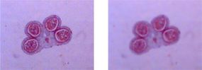
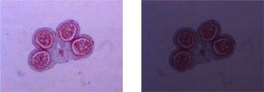
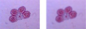
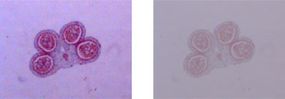
- Contrast - What is the difference in lighting between adjacent areas of the specimen? Contrast is related to the illumination system and can be adjusted by changing the intensity of the light and the diaphragm/pinhole aperture. Also, chemical stains applied to the specimen can enhance contrast.
In the next section, we'll talk about the different types of microscopy.
Advertisement