Types of Microscopy
A major problem in observing specimens under a microscope is that their images do not have much contrast. This is especially true of living things (such as cells), although natural pigments, such as the green in leaves, can provide good contrast. One way to improve contrast is to treat the specimen with colored pigments or dyes that bind to specific structures within the specimen. Different types of microscopy have been developed to improve the contrast in specimens. The specializations are mainly in the illumination systems and the types of light passed through the specimen. For example, a darkfield microscope uses a special condenser to block out most of the bright light and illuminate the specimen with oblique light, much like the moon blocks the light from the sun in a solar eclipse. This optical set-up provides a totally dark background and enhances the contrast of the image to bring out fine details -- bright areas at boundaries within the specimen.
Here are the various types of light microscopy techniques:
Advertisement
- Brightfield - This is the basic microscope configuration (the images seen thus far are all from brightfield microscopes). This technique has very little contrast; in the images you've seen so far, much of the contrast has been provided by staining the specimens.
- Darkfield - This configuration enhances contrast, as mentioned above. See Molecular Expressions: Darkfield Microscopy for details and examples.
- Rheinberg illumination - This set-up is similar to darkfield, but uses a series of filters to produce an "optical staining" of the specimen. See Molecular Expressions: Rheinberg Illumination for details and examples.
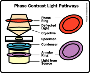
The following techniques use the same basic principle as Rheinberg illumination, achieving different results by using different optical components. The basic idea involves splitting the light beam into two pathways that illuminate the specimen. Light waves that pass through dense structures within the specimen slow down compared to those that pass through less-dense structures. As all of the light waves are collected and transmitted to the eyepiece, they are recombined, so they interfere with each other. The interference patterns provide contrast: They may show dark areas (more dense) on a light background (less dense), or create a type of false three-dimensional (3-D) image.
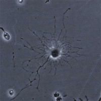
- Phase contrast - This technique is best for looking at living specimens, such as cultured cells.

- Differential interference contrast (DIC) - DIC uses polarizing filters and prisms to separate and recombine the light paths, giving a 3-D appearance to the specimen (DIC is also called Nomarski after the man who invented it). See Molecular Expressions: Differential Interference Contrast Microscopy for details and examples.
- Hoffman modulation contrast - Hoffman modulation contrast is similar to DIC except that it uses plates with small slits in both the axis and the off-axis of the light path to produce two sets of light waves passing through the specimen. Again, a 3-D image is formed. See Molecular Expressions: Hoffman Modulation Contrast Microscopy for details and examples.
- Polarization - The polarized-light microscope uses two polarizers, one on either side of the specimen, positioned perpendicular to each other so that only light that passes through the specimen reaches the eyepiece. Light is polarized in one plane as it passes through the first filter and reaches the specimen. Regularly-spaced, patterned or crystalline portions of the specimen rotate the light that passes through. Some of this rotated light passes through the second polarizing filter, so these regularly spaced areas show up bright against a black background. See Molecular Expressions: Introduction to Polarized Light Microscopy for details and examples.
- Fluorescence - This type of microscope uses high-energy, short-wavelength light (usually ultraviolet) to excite electrons within certain molecules inside a specimen, causing those electrons to shift to higher orbits. When they fall back to their original energy levels, they emit lower-energy, longer-wavelength light (usually in the visible spectrum), which forms the image.
In the next section, we'll discuss fluorescence microscopy in greater detail.
