Hard-wired
The brain is hard-wired with connections, much like a skyscraper or airplane is hard-wired with electrical wiring. In the case of the brain, the connections are made by neurons that link the sensory inputs and motor outputs with centers in the various lobes of the cerebral cortex. There are also linkages between these cortical centers and other parts of the brain.
Several areas of the cerebral cortex have specialized functions:
Advertisement
Parietal lobe -- The parietal lobe receives and processes all somatosensory input from the body (touch, pain).
- Fibers from the spinal cord are distributed by the thalamus to various parts of the parietal lobe.
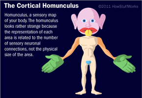
- The connections form a map of the body's surface on the parietal lobe. This map is called a homunculus.
- The rear of the parietal lobe (next to the temporal lobe) has a section called Wernicke's area, which is important for understanding the sensory (auditory and visual) information associated with language. Damage to this area of the brain produces what is called sensory aphasia, in which patients cannot understand language but can still produce sounds.
Frontal lobe -- The frontal lobe is involved in motor skills (including speech) and cognitive functions.
- The motor center of the brain (pre-central gyrus) is located in the rear of the frontal lobe, just in front of the parietal lobe. It receives connections from the somatosensory portion in the parietal lobe and processes and initiates motor functions. Like the homunculus in the parietal lobe, the pre-central gyrus has a motor map of the brain (for details, see A Science Odyssey: You Try It: Probe the Brain Activity).
- An area on the left side of the frontal lobe, called Broca's area, processes language by controlling the muscles that make sounds (mouth, lips and larynx). Damage to this area results in motor aphasia, in which patients can understand language but cannot produce meaningful or appropriate sounds.
- Remaining areas of the frontal lobe perform associative processes (thought, learning, memory).
Occipital lobe -- The occipital lobe receives and processes visual information directly from the eyes and relates this information to the parietal lobe (Wernicke's area) and motor cortex (frontal lobe). One of the things it must do is interpret the upside-down images of the world that are projected onto the retina by the lens of the eye.
Temporal lobe -- The temporal lobe processes auditory information from the ears and relates it to Wernicke's area of the parietal lobe and the motor cortex of the frontal lobe.
- Basal ganglia: Also located within the temporal lobe, the basal ganglia work with the cerebellum to coordinate fine motions, such as fingertip movements.
- Limbic system: Located deep within the temporal lobe, the limbic system is important in emotional behavior and controlling movements of visceral muscles (muscles of the digestive tract and body cavities). The limbic system is comprised of the cingulate gyrus, corpus callosum, mammillary body, olfactory tract, amygdala and hippocampus.
- Hippocampus: The hippocampus is located within the temporal lobe and is important for short-term memory.
- Amygdala: The amygdala is located within the temporal lobe and controls social and sexual behavior and other emotions.
- Insula: The insula influences automatic functions of the brainstem. For example, when you hold your breath, impulses from your insula suppress the medulla's breathing centers. The insula also processes taste information, and separates the temporal and frontal lobes.