Pain Signal Reception
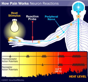
Like normal sensory neurons, nociceptor neurons travel in peripheral sensory nerves. Their cell bodies lie in the dorsal root ganglia of peripheral nerves just inside the spine. As we mentioned, nociceptors sense pain through free nerve endings rather than specialized endings such as those in neurons that sense touch or pressure. However, while normal sensory neurons are myelinated (insulated) and conduct quickly, nociceptor neurons are lightly or non-myelinated and slower. We can divide nociceptors into three classes:
- A δ mechanosensitive receptors -- lightly myelinated, faster conducting neurons that respond to mechanical stimuli (pressure, touch)
- A δ mechanothermal receptors -- lightly myelinated, faster conducting neurons that respond to mechanical stimuli (pressure, touch) and to heat
- Polymodal nociceptors (C fibers) -- unmyelinated, slowly conducting neurons that respond to a variety of stimuli.
Suppose you cut your hand. Several factors contribute to the reception of pain:
Advertisement
- Mechanical stimulation from the sharp object
- Potassium released from the insides of the damaged cells
- Prostaglandins, histamines and bradykinin from immune cells that invade the area during inflammation
- Substance P from nearby nerve fibers
These substances cause action potentials in the nociceptor neurons.
The first thing you may feel when you cut your hand is an intense pain at the moment of the injury. The signal for this pain is conducted rapidly by the A δ-type nociceptors. The pain is followed by a slower, prolonged, dull ache, which is conducted by the slower C-fibers. Using chemical anesthetics, scientists can block one type of neuron and separate the two types of pain.
