How Your Brain Works

The human nervous system
©HowStuffWorks.com

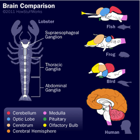
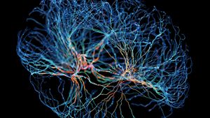
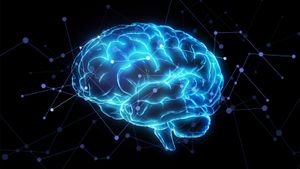
Every animal you can think of -- mammals, birds, reptiles, fish, amphibians -- has a brain. But the human brain is unique. Although it's not the largest, it gives us the power to speak, imagine and problem solve. It is truly an amazing organ.

The brain performs an incredible number of tasks including the following:

Advertisement

  • It controls body temperature, blood pressure, heart rate and breathing.
  • It accepts a flood of information about the world around you from your various senses (seeing, hearing, smelling, tasting and touching).
  • It handles your physical movement when walking, talking, standing or sitting.
  • It lets you think, dream, reason and experience emotions.

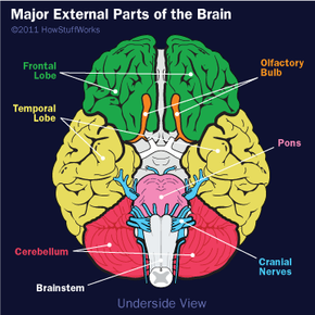
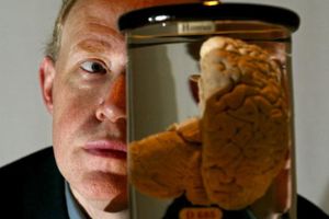
All of these tasks are coordinated, controlled and regulated by an organ that is about the size of a small head of cauliflower.

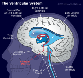
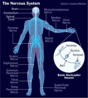
Your brain, spinal cord and peripheral nerves make up a complex, integrated information-processing and control system known as your central nervous system. In tandem, they regulate all the conscious and unconscious facets of your life. The scientific study of the brain and nervous system is called neuroscience or neurobiology. Because the field of neuroscience is so vast -- and the brain and nervous system are so complex -- this article will start with the basics and give you an overview of this complicated organ.

We'll examine the structures of the brain and how each section controls our daily functions, including motor control, visual processing, auditory processing, sensation, learning, memory and emotions.

Neuron Structure

The basic design of a neuron
©HowStuffWorks.com

Your brain is made of approximately 100 billion nerve cells, called neurons. Neurons have the amazing ability to gather and transmit electrochemical signals -- think of them like the gates and wires in a computer.

Neurons share the same characteristics and have the same makeup as other cells, but the electrochemical aspect lets them transmit signals over long distances (up to several feet or a few meters) and send messages to each other.

Advertisement

Neurons have three basic parts:

  • Cell body or soma. This main part has all of the necessary components of the cell, such as the nucleus (which contains DNA), endoplasmic reticulum and ribosomes (for building proteins) and mitochondria (for making energy). If the cell body dies, the neuron dies.
  • Axon . This long, cablelike projection of the cell carries the electrochemical message (nerve impulse or action potential) along the length of the cell. Depending upon the type of neuron, axons can be covered with a thin layer of myelin sheath, like an insulated electrical wire. Myelin is made of fat and protein, and it helps to speed transmission of a nerve impulse down a long axon. Myelinated neurons are typically found in the peripheral nerves (sensory and motor neurons), while non-myelinated neurons are found in the brain and spinal cord.
  • Dendrites or nerve endings. These small, branchlike projections of the cell make connections to other cells and allow the neuron to talk with other cells or perceive the environment. Dendrites can be located on one or both ends of a cell.

Basic Neuron Types

Meet the neurons!
©HowStuffWorks.com

Neurons come in many sizes. For example, a single sensory neuron from your fingertip has an axon that extends the length of your arm, while neurons within the brain may extend only a few millimeters.

They also have different shapes depending on their functions. Motor neurons that control muscle contractions have a cell body on one end, a long axon in the middle and dendrites on the other end. Sensory neurons have dendrites on both ends, connected by a long axon with a cell body in the middle. Interneurons, or associative neurons, carry information between motor and sensory neurons.

Advertisement

These fundamental members of the nervous system also vary with respect to their functions.

  • Sensory neurons carry signals from the outer parts of your body (periphery) into the central nervous system.
  • Motor neurons (motoneurons) carry signals from the central nervous system to the outer parts (muscles, skin, glands) of your body.
  • Interneurons connect various neurons within the brain and spinal cord.

The simplest type of neural pathway is a monosynaptic (single connection) reflex pathway, like the knee-jerk reflex. When the doctor taps the right spot on your knee with a rubber hammer, receptors send a signal into the spinal cord through a sensory neuron. The sensory neuron passes the message to a motor neuron that controls your leg muscles. Nerve impulses travel down the motor neuron and stimulate the appropriate leg muscle to contract. The response is a muscular jerk that happens quickly and does not involve your brain. Humans have lots of hardwired reflexes like this, but as tasks become more complex, the pathway circuitry gets more complicated and the brain gets involved.

Brain Parts

As you proceed up the evolutionary ladder from fish toward humans, check out the changes in the brain. For example, the cerebrum gets bigger, takes up a larger part of the total brain and becomes folded.
©HowStuffWorks.com

The simplest possible creatures have incredibly basic nervous systems made up of nothing but reflex pathways. For example, flatworms and invertebrates don't have centralized brains. They have loose associations of neurons arranged in straightforward reflex pathways. Flatworms have neural nets, or individual neurons linked together that form a net around the entire animal.

Most invertebrates (such as the lobster) have modest "brains" that consist of localized collections of neuronal cell bodies called ganglia. Each ganglion controls sensory and motor functions in its segment through reflex pathways, and the ganglia are linked together to form a simple nervous system. As nervous systems evolved, chains of ganglia evolved into more centralized simple brains.

Advertisement

Brains evolved from ganglia of invertebrates. Regardless of the animal, brains have the following parts:

  • The brain stem, which consists of the medulla (an enlarged portion of the upper spinal cord), pons and midbrain (lower animals have only a medulla). The brain stem controls the reflexes and automatic functions (heart rate, blood pressure), limb movements and visceral functions (digestion, urination).
  • The cerebellum integrates information from the vestibular system that indicates position and movement and uses this data to coordinate limb movements.
  • The hypothalamus and pituitary gland are responsible for visceral functions, body temperature and behavioral responses such as feeding, drinking, sexual response, aggression and pleasure.
  • The cerebrum (also called the cerebral cortex or just the cortex) consists of the cortex, large fiber tracts (corpus callosum) and some deeper structures (basal ganglia, amygdala and hippocampus). It integrates info from all of the sense organs, initiates motor functions, controls emotions and holds memory and thought processes (emotional expression and thinking are more prevalent in higher mammals).

Brains for Instinct

Here we're looking at the underside of the brain, showing the brain stem and cranial nerves.
©HowStuffWorks.com

Lower animals, such as fish, amphibians, reptiles and birds, don't do much "thinking," but instead concern themselves with the everyday business of gathering food, eating, drinking, sleeping, reproducing and defending themselves.

These are instinctual processes [source: National Geographic]. Therefore, their brains are organized along the major centers that control these functions.

Advertisement

We humans perform these functions as well, and so have a "reptilian" brain built into us. That means we have the same parts of the brain found in reptiles, namely the brain stem and the cerebellum.

Ready to learn about the lower brain? We'll discuss that on the next page.

Lower Brain

A peek at the lower brain
©HowStuffWorks.com

The basic lower brain consists of the spinal cord, brain stem and diencephalon (the cerebellum and cortex are also present, but will be discussed in later sections). In turn, the brain stem comprises the medulla, pons, midbrain, hypothalamus and thalamus [source: Health Pages].

Within each of these structures are centers of neuronal cell bodies, called nuclei, which are specialized for particular functions (breathing, heart-rate regulation, sleep):

Advertisement

  • Medulla -- The medulla contains nuclei for regulating blood pressure and breathing, as well as nuclei for relaying information from the sense organs that comes in from the cranial nerves. It's also the most ancient part of the brain.
  • Pons -- The pons contains nuclei that relay movement and position information from the cerebellum to the cortex. It also contains nuclei that are involved in breathing, taste and sleep, and physically connects medulla to the midbrain.
  • Midbrain -- The midbrain contains nuclei that link the various sections of the brain involved in motor functions (cerebellum, basal ganglia, cerebral cortex), eye movements and auditory control. One portion, called the substantia nigra, is involved in voluntary movements; when it does not function, you have the tremored movements of Parkinson's disease.
  • Thalamus -- The thalamus relays incoming sensory pathways to appropriate areas of the cortex, determines which sensory information actually reaches consciousness and participates in motor-information exchange between the cerebellum, basal ganglia and cortex.
  • Hypothalamus -- The hypothalamus contains nuclei that control hormonal secretions from the pituitary gland. These centers govern sexual reproduction, eating, drinking, growth, and maternal behavior such as lactation (milk-production in mammals). The hypothalamus is also involved in almost all aspects of behavior, including your biological "clock," which is linked to the daily light-dark cycle (circadian rhythms).

Balancing Act

The cerebellum, also known as the "little brain" because it's folded into many lobes, lies above and behind the pons. As the second biggest area of the brain, it receives sensory input from the spinal cord, motor input from the cortex and basal ganglia, and position information from the vestibular system.

The "little brain" then integrates this information and influences outgoing motor pathways from the brain to coordinate movements. To demonstrate this, reach out and touch a point in front of you, such as the computer monitor -- your hand makes one smooth motion. If your cerebellum were damaged, that same motion would be very jerky, as your cortex initiated a series of small muscle contractions to home in on the target point. The cerebellum may also be involved in language (fine muscle contractions of the lips and larynx), as well as other cognitive functions.

Advertisement

Higher Brains

External parts of the human brain
©HowStuffWorks.com

The cerebrum is the largest part of the human brain. It contains all of the centers that receive and interpret sensory information, initiate movement, analyze information, reason and experience emotions. The centers for these tasks are located in different parts of the cerebral cortex, which is the outside layer of the cerebellum and is comprised of gray matter. The inside is made up of white matter.

Major Parts of the Cerebral Cortex

Advertisement

The cortex dominates the exterior surface of the brain. The surface area of the brain is about 233 to 465 square inches (1,500 to 2,000 cm2), which is about the size of one to two pages of a newspaper. To fit this surface area within the skull, the cortex is folded, forming folds (gyri) and grooves (sulci). Several large sulci divide the cerebral cortex into various lobes: the frontal lobe, parietal lobe, occipital lobe and temporal lobe. Each lobe has a different function.

Get to know the interior of your brain a bit better.
©HowStuffWorks.com

When viewed from above, a large groove (interhemispheric fissure) separates the brain into left and right halves. The halves talk to each other through a tract of white-matter fibers called the corpus callosum. Also, the right and left temporal lobes communicate through another tract of fibers near the rear of the brain called the anterior commissure.

If you look at a cutaway view of the brain, you see that the cortical area above the corpus callosum is divided by a groove. This groove is called the cingulate sulcus. The area between that groove and the corpus callosum is called the cingulate gyrus, also referred to as the limbic system or limbic lobe. Deep within the cerebrum are the basal ganglia, amygdala and hippocampus.

This ends our tour of the major structures of the cortex. Now, let's see what those structures do.

Hard-wired

Sure, this homunculus looks rather strange, but that's because the representation of each area is related to the number of sensory neuronal connections, not its physical size.
©HowStuffWorks.com

The brain is hard-wired with connections, much like a skyscraper or airplane is hard-wired with electrical wiring. In the case of the brain, the connections are made by neurons that link the sensory inputs and motor outputs with centers in the various lobes of the cerebral cortex. There are also linkages between these cortical centers and other parts of the brain.

Several areas of the cerebral cortex have specialized functions:

Advertisement

Parietal lobe -- The parietal lobe receives and processes all somatosensory input from the body (touch, pain).

  • Fibers from the spinal cord are distributed by the thalamus to various parts of the parietal lobe.
  • The connections form a map of the body's surface on the parietal lobe. This map is called a homunculus.
  • The rear of the parietal lobe (next to the temporal lobe) has a section called Wernicke's area, which is important for understanding the sensory (auditory and visual) information associated with language. Damage to this area of the brain produces what is called sensory aphasia, in which patients cannot understand language but can still produce sounds.

Frontal lobe -- The frontal lobe is involved in motor skills (including speech) and cognitive functions.

  • The motor center of the brain (pre-central gyrus) is located in the rear of the frontal lobe, just in front of the parietal lobe. It receives connections from the somatosensory portion in the parietal lobe and processes and initiates motor functions. Like the homunculus in the parietal lobe, the pre-central gyrus has a motor map of the brain (for details, see A Science Odyssey: You Try It: Probe the Brain Activity).
  • An area on the left side of the frontal lobe, called Broca's area, processes language by controlling the muscles that make sounds (mouth, lips and larynx). Damage to this area results in motor aphasia, in which patients can understand language but cannot produce meaningful or appropriate sounds.
  • Remaining areas of the frontal lobe perform associative processes (thought, learning, memory).

Occipital lobe -- The occipital lobe receives and processes visual information directly from the eyes and relates this information to the parietal lobe (Wernicke's area) and motor cortex (frontal lobe). One of the things it must do is interpret the upside-down images of the world that are projected onto the retina by the lens of the eye.

Temporal lobe -- The temporal lobe processes auditory information from the ears and relates it to Wernicke's area of the parietal lobe and the motor cortex of the frontal lobe.

  • Basal ganglia: Also located within the temporal lobe, the basal ganglia work with the cerebellum to coordinate fine motions, such as fingertip movements.
  • Limbic system: Located deep within the temporal lobe, the limbic system is important in emotional behavior and controlling movements of visceral muscles (muscles of the digestive tract and body cavities). The limbic system is comprised of the cingulate gyrus, corpus callosum, mammillary body, olfactory tract, amygdala and hippocampus.
  • Hippocampus: The hippocampus is located within the temporal lobe and is important for short-term memory.
  • Amygdala: The amygdala is located within the temporal lobe and controls social and sexual behavior and other emotions.
  • Insula: The insula influences automatic functions of the brainstem. For example, when you hold your breath, impulses from your insula suppress the medulla's breathing centers. The insula also processes taste information, and separates the temporal and frontal lobes.

Water on the Brain

The human brain's ventricular system
©HowStuffWorks.com

Your brain and spinal cord are covered by a series of tough membranes called meninges, which protect these organs from rubbing against the bones of the skull and spine.

For further protection, the brain and spinal cord "float" in a sea of cerebrospinal fluid within the skull and spine. This cushioning fluid is produced by the choroid plexus tissue, which is located within the brain, and flows through a series of cavities (ventricles) out of the brain and down along the spinal cord. The cerebrospinal fluid is kept separate from the blood supply by the blood-brain barrier.

Advertisement

As you can see, your brain is a complex, highly organized organ that governs everything you do. Now that you are familiar with the anatomy of the brain, keep reading for more articles on how it works.

Lots More Information

Related Articles

  • Bryn Mawr University. "Brain Structures and their Functions." June 3, 2005. (June 13, 2011). http://serendip.brynmawr.edu/bb/kinser/Structure1.html
  • Children's Hospital Boston. "The Neuron." 2007. (June 13, 2011). http://www.childrenshospital.org/research/_neuron/index.html
  • Healthpages.org. "Anatomy of the Brain." Feb. 28, 2011. (June 15, 2011). http://healthpages.org/anatomy-function/brain-anatomy/
  • McGill University. "The Brain From Top To Bottom." (June 14, 2011).http://thebrain.mcgill.ca/flash/d/d_05/d_05_cr/d_05_cr_her/d_05_cr_her.html
  • National Geographic. "Command Central." 1996-2011. (June 14, 2011). http://science.nationalgeographic.com/science/health-and-human-body/human-body/brain-article/
  • The Science Museum. "What makes the human brain unique." Sciencemuseum.org. (June 13, 2011). http://www.sciencemuseum.org.uk/WhoAmI/FindOutMore/Yourbrain/Howdoesyourbrainwork/Whatarethepartsofyourbrain/Whatmakesthehumanbrainunique.aspx
  • Washington University, Neuroscience for Kids. "Types of Neurons." 1996-2011. (June 13, 2011). http://faculty.washington.edu/chudler/cells.html