How X-rays Work

By: Tom Harris  | 
x-rays of King George and Queen Mary
This X-ray is of the hands of Queen Mary and King George and was taken just a year after the monumental and accidental discovery of X-rays in 1895. Science & Society Picture Library/SSPL via Getty Images

As with many of mankind's monumental discoveries, X-ray technology was invented completely by accident. In 1895, a German physicist named Wilhelm Roentgen made the discovery while experimenting with electron beams in a gas discharge tube. Roentgen noticed that a fluorescent screen in his lab started to glow when the electron beam was turned on. This response in itself wasn't so surprising -- fluorescent material normally glows in reaction to electromagnetic radiation -- but Roentgen's tube was surrounded by heavy black cardboard. Roentgen assumed this would have blocked most of the radiation.

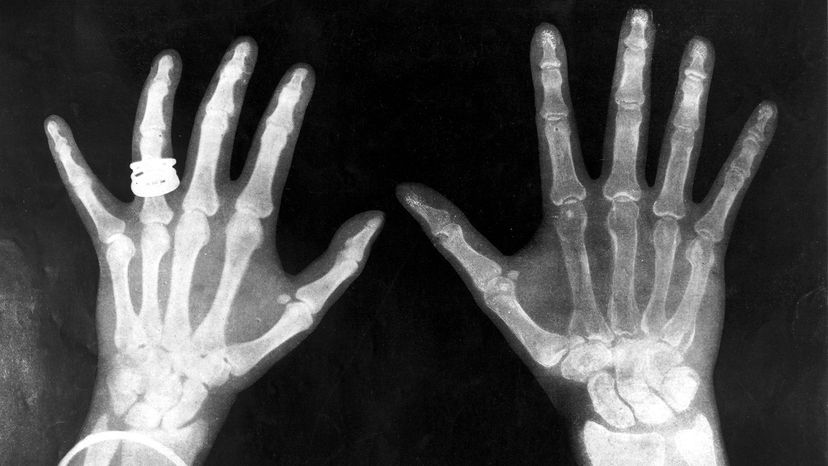
Roentgen placed various objects between the tube and the screen, and the screen still glowed. Finally, he put his hand in front of the tube, and saw the silhouette of his bones projected onto the fluorescent screen. Immediately after discovering X-rays themselves, he had discovered their most beneficial application.

Advertisement

Roentgen's remarkable discovery precipitated one of the most important medical advancements in human history. X-ray technology lets doctors see straight through human tissue to examine broken bones, cavities and swallowed objects with extraordinary ease. Modified X-ray procedures can be used to examine softer tissue, such as the lungs, blood vessels or the intestines.

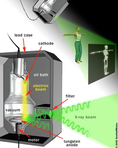
In this article, we'll find out exactly how X-rays machines pull off this incredible trick. As it turns out, the basic process is really very simple.

Advertisement

What's an X-Ray?

X-rays are basically the same thing as visible light rays. Both are wavelike forms of electromagnetic energy carried by particles called photons (see How Light Works for details). The difference between X-rays and visible light rays is the energy level of the individual photons. This is also expressed as the wavelength of the rays.

Our eyes are sensitive to the particular wavelength of visible light, but not to the shorter wavelength of higher energy X-ray waves or the longer wavelength of the lower energy radio waves.

Advertisement

Visible light photons and X-ray photons are both produced by the movement of electrons in atoms. Electrons occupy different energy levels, or orbitals, around an atom's nucleus. When an electron drops to a lower orbital, it needs to release some energy -- it releases the extra energy in the form of a photon. The energy level of the photon depends on how far the electron dropped between orbitals. (See this page for a detailed description of this process.)

When a photon collides with another atom, the atom may absorb the photon's energy by boosting an electron to a higher level. For this to happen, the energy level of the photon has to match the energy difference between the two electron positions. If not, the photon can't shift electrons between orbitals.

The atoms that make up your body tissue absorb visible light photons very well. The energy level of the photon fits with various energy differences between electron positions. Radio waves don't have enough energy to move electrons between orbitals in larger atoms, so they pass through most stuff. X-ray photons also pass through most things, but for the opposite reason: They have too much energy.

They can, however, knock an electron away from an atom altogether. Some of the energy from the X-ray photon works to separate the electron from the atom, and the rest sends the electron flying through space. A larger atom is more likely to absorb an X-ray photon in this way, because larger atoms have greater energy differences between orbitals -- the energy level more closely matches the energy of the photon. Smaller atoms, where the electron orbitals are separated by relatively low jumps in energy, are less likely to absorb X-ray photons.

The soft tissue in your body is composed of smaller atoms, and so does not absorb X-ray photons particularly well. The calcium atoms that make up your bones are much larger, so they are better at absorbing X-ray photons.

In the next section, we'll see how X-ray machines put this effect to work.

Advertisement

The X-Ray Machine

The heart of an X-ray machine is an electrode pair -- a cathode and an anode -- that sits inside a glass vacuum tube. The cathode is a heated filament, like you might find in an older fluorescent lamp. The machine passes current through the filament, heating it up. The heat sputters electrons off of the filament surface. The positively-charged anode, a flat disc made of tungsten, draws the electrons across the tube.

X-ray machines seem to do the impossible: They see straight through clothing, flesh and even metal thanks to some very cool scientific principles at work. Find out how X-ray machines see straight to your bones.

The voltage difference between the cathode and anode is extremely high, so the electrons fly through the tube with a great deal of force. When a speeding electron collides with a tungsten atom, it knocks loose an electron in one of the atom's lower orbitals. An electron in a higher orbital immediately falls to the lower energy level, releasing its extra energy in the form of a photon. It's a big drop, so the photon has a high energy level -- it is an X-ray photon.

Advertisement

Free electrons can also generate photons without hitting an atom. An atom's nucleus may attract a speeding electron just enough to alter its course. Like a comet whipping around the sun, the electron slows down and changes direction as it speeds past the atom. This "braking" action causes the electron to emit excess energy in the form of an X-ray photon.

The high-impact collisions involved in X-ray production generate a lot of heat. A motor rotates the anode to keep it from melting (the electron beam isn't always focused on the same area). A cool oil bath surrounding the envelope also absorbs heat.

The entire mechanism is surrounded by a thick lead shield. This keeps the X-rays from escaping in all directions. A small window in the shield lets some of the X-ray photons escape in a narrow beam. The beam passes through a series of filters on its way to the patient.

A camera on the other side of the patient records the pattern of X-ray light that passes all the way through the patient's body. The X-ray camera uses the same film technology as an ordinary camera, but X-ray light sets off the chemical reaction instead of visible light. (See How Photographic Film Works to learn about this process.)

Generally, doctors keep the film image as a negative. That is, the areas that are exposed to more light appear darker and the areas that are exposed to less light appear lighter. Hard material, such as bone, appears white, and softer material appears black or gray. Doctors can bring different materials into focus by varying the intensity of the X-ray beam.

Advertisement

Are X-Rays Bad for You?

X-rays are a wonderful addition to the world of medicine; they let doctors peer inside a patient without any surgery at all. It's much easier and safer to look at a broken bone using X-rays than it is to open a patient up.

But X-rays can also be harmful. In the early days of X-ray science, a lot of doctors would expose patients and themselves to the beams for long periods of time. Eventually, doctors and patients started developing radiation sickness, and the medical community knew something was wrong.

Advertisement

The problem is that X-rays are a form of ionizing radiation. When normal light hits an atom, it can't change the atom in any significant way. But when an X-ray hits an atom, it can knock electrons off the atom to create an ion, an electrically charged atom. Free electrons then collide with other atoms to create more ions.

An ion's electrical charge can lead to unnatural chemical reactions inside cells. Among other things, the charge can break DNA chains. A cell with a broken strand of DNA will either die or the DNA will develop a mutation. If a lot of cells die, the body can develop various diseases. If the DNA mutates, a cell may become cancerous, and this cancer may spread. If the mutation is in a sperm or an egg cell, it may lead to birth defects. Because of all these risks, doctors use X-rays sparingly today.

Even with these risks, X-ray scanning is still a safer option than surgery. X-ray machines are an invaluable tool in medicine, as well as an asset in security and scientific research. They are truly one of the most useful inventions of all time.

For more information on X-rays and X-ray machines, check out the links that follow.

Advertisement

Frequently Asked Questions

What were X-rays used for?
X-rays are commonly used in medicine for imaging internal structures to diagnose conditions such as fractures, tumors and dental issues. Additionally, X-rays find applications in industrial inspections, security screening, scientific research and cancer treatment through radiotherapy.
What are X-rays made of?
X-rays are a form of electromagnetic radiation, similar to visible light but with much higher energy. They consist of photons, which are particles without mass or charge, and their energy allows them to penetrate materials and create images of internal structures in various applications such as medical imaging and industrial inspections.
Does arthritis show up on X-rays?
Arthritis can be visible on X-rays as it often leads to changes in the structure of joints, including narrowing of joint spaces, bone spurs, and alterations in bone density. However, early stages of arthritis may not always be clearly evident on X-rays, and other imaging methods or clinical assessments may be needed for an accurate diagnosis.

Advertisement

Loading...