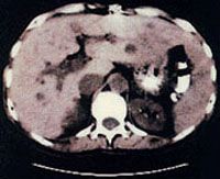
Computerized axial tomography (CAT) scan machines produce X-rays, a powerful form of electromagnetic energy. X-ray photons are basically the same thing as visible light photons, but they have much more energy. This higher energy level allows X-ray beams to pass straight through most of the soft material in the human body. (See How X-Rays Work to find how X-rays do this, as well as how X-ray machines produce X-ray photons).
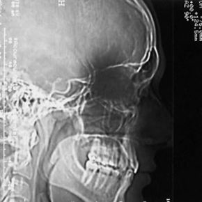
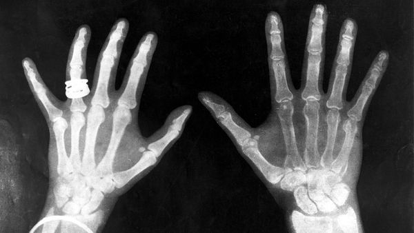
A conventional X-ray image is basically a shadow: You shine a "light" on one side of the body, and a piece of film on the other side registers the silhouette of the bones.
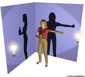
Shadows give you an incomplete picture of an object's shape. Imagine you are standing in front of a wall, holding a pineapple against your chest with your right hand and a banana out to your side with your left hand. Your friend is looking only at the wall, not at you. If there's a lamp in front of you, your friend will see the outline of you holding the banana, but not the pineapple -- the shadow of your torso blocks the pineapple. If the lamp is to your left, your friend will see the outline of the pineapple, but not the banana.
The same thing happens in a conventional X-ray image. If a larger bone is directly between the X-ray machine and a smaller bone, the larger bone may cover the smaller bone on the film. In order to see the smaller bone, you would have to turn your body or move the X-ray machine.
In order to know that you are holding a pineapple and a banana, your friend would have to see your shadow in both positions and form a complete mental image. This is the basic idea of computer aided tomography. In a CAT scan machine, the X-ray beam moves all around the patient, scanning from hundreds of different angles. The computer takes all this information and puts together a 3-D image of the body.