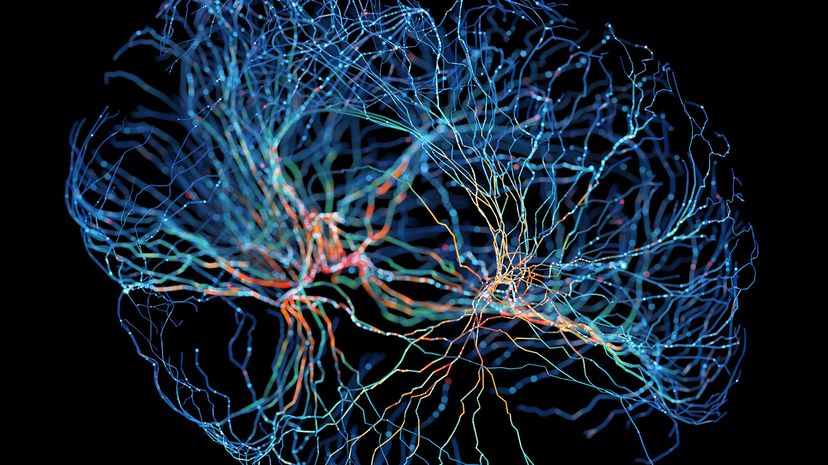
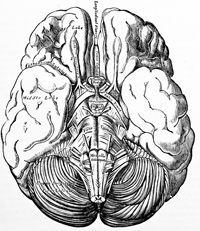
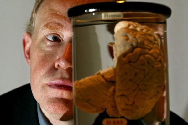
Why would scientists take on the arduous task of brain mapping? The answer is simple, says Lichtman: to understand our brains more intimately.
We have never seen a diagram of how all of the neurons in the brain connect. As Jeff Lichtman puts it, "A lot of our thinking about the brain is based on incomplete knowledge of what is actually there. So we would like to see what is actually there."
The brain's wiring diagram may help us better understand how we learn and adapt, says Lichtman:
"We start out being less well-adapted to our environment than any other animal. By the time we're adults, we can use tools that our genetic heritage couldn't possibly have taught our nervous system to use — like iPods. No other animal can do that. During our development, we must wire ourselves to [be able to] use these machines."
Brain mapping is also of practical use to doctors. Neurosurgeons use brain mapping to plan safer surgeries. One treatment for epilepsy, for example, removes the affected part of the brain.
Using functional MRI and EEG, surgeons can locate the seizure center in a patient's brain — as well as areas that are active during speaking and moving — down to the millimeter. These images tell doctors what to leave and what to cut out.
Brain imaging is not only used in treatment. It is used to diagnose neurodegenerative diseases like Parkinson's and Alzheimer's [source: Wilson]. Using tagging techniques like PET, doctors look for drops in certain brain chemicals, or they may use MRI to examine shrinkages in areas show tissue loss.
Over time, doctors can map what the brain looks like as diseases progress or as treatments work [source: Institute for Neurodegenerative Disorders].
Developmental disorders like autism may have a structural basis in the brain. Lichtman points out that autism is thought to involve a series of unique connections between neurons. By applying Brainbow to a mouse with autism, researchers might see the wiring diagram evolve to find out how, when and if the wiring differs from a mouse without autism.
Scientists have also sought to illustrate the effects of various psychiatric disorders in the brain, with some success. Brain imaging on these patients revealed structural abnormalities. For example, structural MRI has shown that schizophrenic patients lose matter in the temporal and prefrontal cortex over time [Source: Thompson et al.].
Panic disorder, bipolar disorder, depression, anxiety, eating disorders and more are being examined using different brain imaging techniques, but how do we interpret scientists' findings? More importantly, where can we see them?
Doctors and scientists have learned more from brain mapping than this article can cover. Here are two highlights:
Sarcasm
We detect sarcasm using a brain region called the right parahippocampal gyrus. Researchers discovered this using functional MRI on patients with deterioration in that region and have lost all sense of sarcasm [source: Hurley].
Consciousness
According to Rodolfo Llinas of New York University, we can divide the brain into a synchronization center deep in the brain and neuron loops that give us higher thought.
We feel conscious when the center keeps the loops working in harmony. But if either part is damaged, we may lose some or all consciousness, says Nicholas Schiff at Cornell University's Weill Medical College.
This may explain why patients with no outward signs of consciousness for years may show completely normal brain activity in response to a familiar voice. They have loops working in isolation, like the neural networks that process language [source: Zimmer].
