Different Types of Ultrasound
The ultrasound that we have described so far presents a two-dimensional image, or "slice," of a three-dimensional object (fetus, organ). Two other types of ultrasound are currently in use, 3-D ultrasound imaging and Doppler ultrasound.
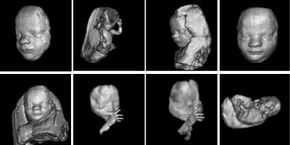
Ultrasound machines capable of three-dimensional imaging have been developed. In these machines, several two-dimensional images are acquired by moving the probes across the body surface or rotating inserted probes. The two-dimensional scans are then combined by specialized computer software to form 3-D images.
Advertisement
3-D imaging allows you to get a better look at the organ being examined and is best used for:
- Early detection of cancerous and benign tumors (examining the prostate gland for early detection of tumors, looking for masses in the colon and rectum, detecting breast lesions for possible biopsies).
- Visualizing a fetus to assess its development, especially for observing abnormal development of the face and limbs.
- Visualizing blood flow in various organs or a fetus.
Doppler ultrasound is based upon the Doppler Effect. When the object reflecting the ultrasound waves is moving, it changes the frequency of the echoes, creating a higher frequency if it is moving toward the probe and a lower frequency if it is moving away from the probe. How much the frequency is changed depends upon how fast the object is moving. Doppler ultrasound measures the change in frequency of the echoes to calculate how fast an object is moving. Doppler ultrasound has been used mostly to measure the rate of blood flow through the heart and major arteries.