Operating a Scanning Electron Microscope
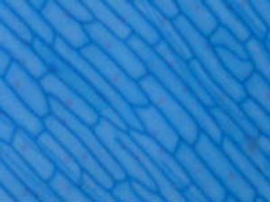
Before researchers can take their first SEM image of, say, a mosquito, they have to prepare the specimen. Because SEMs, unlike optical microscopes, operate in a vacuum and rely on electric fields to work, sample preparation can be a complicated process. Researchers start by cleaning it of any dust or debris. Once clean, it's ready to be mounted in the SEM if the specimen is fairly conductive. Otherwise, it's coated in a conductive material like gold or platinum through a process called sputter coating before it's ready for viewing. Sputter coating allows a sample to be grounded, preventing it from being damaged by the electron beam.
Since specimens placed in the microscopes also are subject to a vacuum, they sometimes undergo additional preparation to ensure that they hold up under such extreme conditions. Biological samples, for instance, are typically dehydrated before being placed in an SEM. Otherwise, the low atmospheric pressure of a vacuum would cause the water in biological samples to evaporate quickly, destroying the sample in the process. Other specimens are frozen before they are examined, and still others are chemically treated so that they survive the magnification process.
Advertisement
Researchers, like photographers, have a variety of controls over the images they produce. The magnification, focus, contrast and brightness of an image are all at the fingertips of the operator of an SEM. While some models have dedicated hardware for these settings, the more recent integration of computerized controls has both lowered the cost of SEMs and simplified their operation.
Finally, make sure to observe some safety precautions when operating the instrument. In the process of scanning specimens, SEMs generate small levels of radiation in the form of X-rays as electrons beneath the surface of a specimen are dislodged and replaced by other electrons. While X-rays are inherently dangerous to humans, you shouldn't be too worried about operating an SEM. Most of the instruments have a highly isolated specimen chamber, designed to keep out electrical and magnetic interference, so any X-rays generated in the magnification process shouldn't pose a threat to the operator. Still, researchers should make sure to observe any safety precautions concerning the operation of the SEMs at their institution.
Related HowStuffWorks Articles
More Great Links
Sources
- Breton, Bernie C. "The Early History and Development of The Scanning Electron Microscope." Cambridge University. Oct. 26, 2006. (April 10, 2009) http://www2.eng.cam.ac.uk/~bcb/history.htm
- Derene, Glenn. "World's Smallest Electron Microscope (for Less than a Bimmer!)." Popular Mechanics. July 6, 2007. (April 10, 2009)http://www.popularmechanics.com/blogs/technology_news/4218957.html
- Grice, Gordon. "Invisible Clues." Popular Science. Sept. 27, 2002. (April 10, 2009)https://www.popsci.com/scitech/article/2002-09/invisible-clues
- Iowa State University. "Understanding how the SEM works and how to use it on a college level." (April 10, 2009)http://mse.iastate.edu/microscopy/college.html
- JEOL, Ltd. "New Scanning Electron Microscope Capable of Observing Cells in Solution." Dec. 8, 2008. (April 10, 2009)http://www.jeol.com/Portals/0/Press/Atmospheric_SEM_English.pdf
- Michigan State University. "Scanning Electron Microscope (SEM) Introduction." (April 10, 2009)http://www.chems.msu.edu/curr.stud/mse.sops/sem.intro.htm
- Physics World. "Beating the diffraction limit." Sept. 1, 2001. (April 10, 2009)http://physicsworld.com/cws/article/print/22
- Scientific Blogging. "The No-Stick Feet Of Mirid Bugs." Aug. 8, 2008. (April 15, 2008)http://www.scientificblogging.com/news_releases/the_no_stick_feet_of_mirid_bugs
- Science Daily. "New Electron Microscope Identifies Individual Color-Coded Atoms." Feb. 22, 2008. (April 10, 2009)http://www.sciencedaily.com/releases/2008/02/080221153725.htm
- Swap, Susan. "Scanning Electron Microscopy." University of Wyoming. April 7, 2009. (April 10, 2009)http://serc.carleton.edu/research_education/geochemsheets/techniques/SEM.html
- University of Nebraska-Lincoln. "What are Electron Microscopes?" (April 10, 2009)http://www.unl.edu/CMRAcfem/em.htm
- University of Wisconsin. "Preparing samples for scanning electron microscopy." (April 10, 2009)http://www.ansci.wisc.edu/facstaff/Faculty/pages/albrecht/albrecht_web/Programs/microscopy/prep.html